Chapter 6: Blood composition
The primary function of blood is to deliver oxygen and nutrients to and remove waste from the body’s cells. However, it is also responsible for transport of hormones, protection (clotting and immune defence), distribution of heat and maintenance of homeostasis (pH and water balance). To carry out these wide-ranging functions, blood has a necessarily complex composition.
Blood has two main components, plasma (blood fluid) and formed elements (cells and platelets). You quickly see these main components by performing a haemocrit test. In this test whole blood is centrifuged to pellet the formed elements at the bottom of the tube, leaving the plasma at the top of the tube. A haemocrit is normally conducted as part of a complete blood count and used to determine the volume percentage of erythrocytes in the blood. For a normal haemocrit the contribution of erythrocytes is about 45% but this varies depending on a number of factors including sex, age, pregnancy and even the altitude you live!
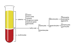
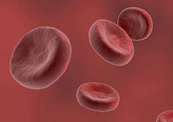
Erythrocytes are the dominant cell type; they comprise 99% of the cells in blood. In a haemocrit you will also see a thin layer of buff-coloured cells above the erythrocytes (buffy coat) which contains most of the leukocytes and thrombocytes (platelets).
How big are erythrocytes?
Estimating cell size
We can estimate cell size by viewing cells under a microscope. The circle of light you see through the ocular lens is known as the field of view. You can determine the width of the field of view by using a stage micrometer, which is a microscope slide that has a scale etched into its surface. A haemocytometer (see the next section) can be used for crude estimates.
Once you have an idea of the width of the field of view you can estimate how many cells laid end to end it would take to equal the diameter of the field of view.

