6.1 Counting cells

The device shown in the next figure is used for accurately determining cell concentrations. It is known as a haemocytometer, since it was originally designed for performing blood cell counts. However, it is also used for cell culture, microbiology or any application where it is necessary to determine the number of cells per unit volume of a suspension.
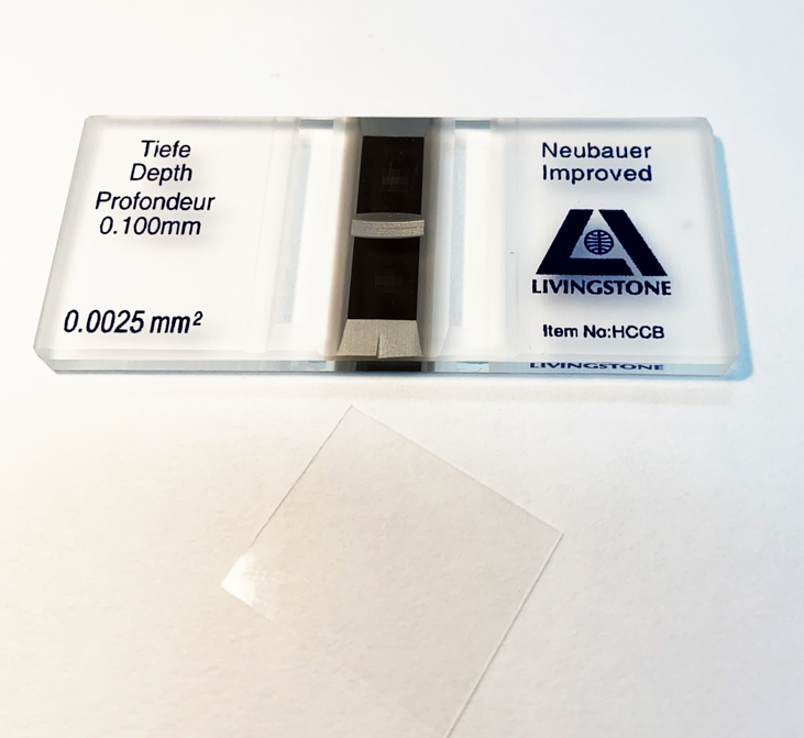
A haemocytometer is a modified and calibrated microscope slide which has two coverslip supports precisely raised above a cell counting surface inscribed with a precise grid, as shown in the next figure. This creates a defined volume for counting cells in suspension, which are pipetted into the counting chamber.
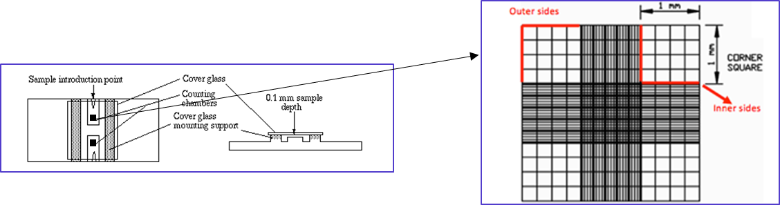
Care is taken to dilute the cell suspension, so it is not so crowded it is impossible to count and also so that the cells are uniformly distributed. Often a dye is added to the cells which distinguishes dead cells from viable cells. The cell suspension is pipetted slowly into the counting chamber, which fills by capillary action. For most mammalian cell suspensions, the large corner squares (1 mm × 1 mm) are used for counting but smaller squares may be used for smaller cells such as erythrocytes or yeast. The cells are counted while viewing the haemocytometer under a microscope. Counting is done systematically to avoid bias. For instance, cells that overlap a line count as ‘in’ if they overlap the top or right outer line, and ‘out’ if they overlap the bottom or left outer line.
Calculating cell concentrations from a haemocytometer
A typical depth for a haemocytometer counting chamber is 0.1 mm. Therefore, the volume defined by one of the 1 mm outer squares is:
height x width x depth = volume
1 mm × 1 mm × 0.1 mm = 0.1 mm3
(there are 1,000 mm3 in 1 cm3 and 1 cm3 = 1 ml)
Therefore, 0.1 mm3 = 1 ml/1,000 mm3 x 0.1 mm3 = 0.0001 ml = 0.1 µl
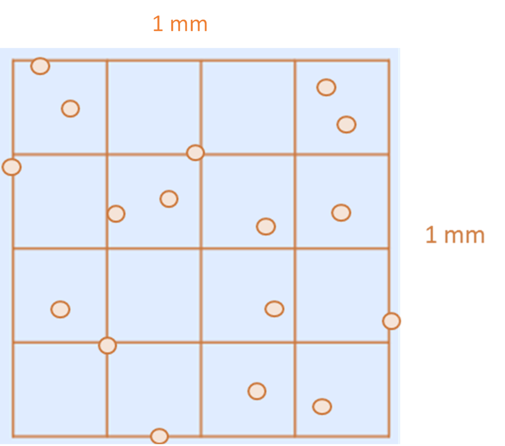
In the 1 mm × 1 mm square above, there are 15 cells (counting to avoid bias). Assuming this is representative of the cell suspension they were aliquoted from; this means that cell suspension contains:
15 cells per 0.1 µl
which is 150 cells per µl
which is 150,000 cells per ml.
