1.3 The Brain
Following the introduction to neurons, and how they are connected and communicate with each other, in the previous topic, we will now turn our attention to how these neurons make up our brain and nervous system. We will focus on those areas that are important for understanding people’s behaviours in conflict, including those structures that are particularly involved in cognition and emotions.
Our brain, together with our spinal cord, makes up our central nervous system. The spinal cord is a long cylinder of nerve tissues that extends from the brain. Understanding the specific areas and functions of the spinal cord is less important for analysing people’s conflict behaviours, which is why we will not focus on this topic here but go straight to the brain.

Watch this video as an introduction to the human brain [12:33]:
As you may have just heard in the video, understanding our brains is critical to understanding our minds (“the mind is what the brain does”), which is again central to this eBook.
Scientists have divided the brain in multiple different ways. To support your understanding of the various brain regions, we will first distinguish between the brain stem (consisting of the hindbrain and the midbrain) and the forebrain below.
The Brainstem
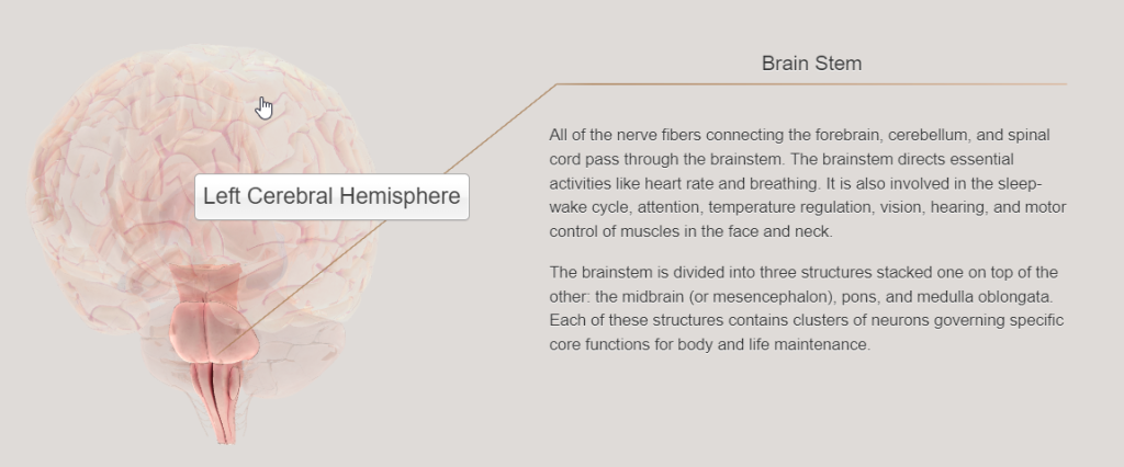
The brainstem is involved in the regulation of vital automatic functions, like breathing and regulating body temperature (Gluck et al., 2020). It comprises several structures that connect the brain with the spinal cord, inducing the medulla, the pons, the cerebellum, as well as the midbrain.
The medulla is located right above the spinal cord and all information passing to and from the higher brain structures to our body must pass through the medulla. The medulla is involved in vital functions like breathing, heart rate and blood pressure, as well as in consciousness, movement and pain.
Above the medulla is the pons, which is involved in processes like sound perception, motor control of eyes, head position and movement. The pons also plays a role in mood, arousal, aggression, appetite, and sleep.
The cerebellum is located at the back of the brain stem. It contains more neurons than the rest of the brain combined. The cerebellum has been found to contribute to motor coordination, the encoding and retrieval of skill memory, executive functions, emotional processing, learning and social awareness. People with autism frequently have abnormal development of the cerebellum and may thus struggle with the processing of emotions and social cues. The cerebellum is also the brain structure that is first affected by alcohol consumption, impacting speech and balance.
The midbrain fulfils several functions, including the communication of information between the spinal cord and the cerebellum. Another function of the midbrain that may be relevant for people in conflict and conflict resolution is that of arousal; after all, emotions experienced by people in conflict typically involve high arousal. One important area of the midbrain is the PAG (which is short for periaqueductal grey). While this area is mainly involved in pain reduction, it also plays an important role in the regulation of the heart rate, blood pressure, autonomic processes, as well as fearful and defensive reactions, which are all implicated in the experience of certain emotions (we will look at these in more detail in Chapter 3). Another structure of the midbrain that may be of interest to neuroscience research on people in conflict is the substantia nigra. This structure is one of the sources for the neurochemical dopamine, which is associated with motivation and reward. You will learn more about dopamine and other neurotransmitters later in this chapter.
The Forebrain
The forebrain contains many structures that play a major role in cognition and the experience of emotions, and is therefore particularly relevant for this eBook. We will look at some of these structures in more detail below.
Cortex
The cerebral cortex is the outer covering of the brain and the largest structure of the brain (Freberg, 2019; Gluck et al., 2020). It is commonly divided into lobes, which are involved in many different functions.
Check out the following video to learn more about these lobes [1:59]:
Before we look at the various lobes in more detail, we will briefly consider some differences between the left and right cerebral hemispheres (called hemispheric asymmetry). For example, two brain regions that are critical for speech production (the Broca’s area and the Wernicke’s area) are located in the left hemisphere of the brain of most right-handed people. For those who are left-handed, the area could be located in the right hemisphere.
Extension:
For an interesting story of a neuroanatomist who survived a stroke and blood clot in her left brain and who discusses, from personal experience, the differences between left-brain and right-brain functions, please watch the following video [20:12]:
Some hemispheric asymmetries discussed in the neuroscience literature may be relevant for understanding and supporting people in conflict. According to Freberg (2019), the right hemisphere is viewed as the more emotional hemisphere and is thought to play a larger role in processing emotions, the perception of emotions in others’ faces, and the expression of emotions. The right side of our brain controls the left side of our face, which is generally more expressive than the right side (this may vary in left-handed people) (Freberg, 2019). Research has found that the meaning of emotion-related words are processed faster by the left hemisphere of the brain, while the identification of emotional tone is more accurately processed by the right hemisphere. The left hemisphere is generally associated with “approach” behaviour, while the right hemisphere is generally associated with “avoidance” (Fetterman et al., 2013; Freberg, 2019). We will consider some of these phenomena in Chapter 3, which focuses on emotions.
Please note that hemispheric asymmetry does not relate to the alleged “creative right versus analytical left brain” myth, which is not supported by neuroscience research (Nielsen et al., 2013).
Extension:
If you want to learn more about the left-brain vs right-brain myth, how it came about and how it is not supported by neuroscience research, please read this blog post by Sarah McKay.
The Four Lobes of the Cerebral Hemispheres
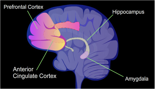
The frontal lobe is of great interest to this eBook since it participates in many higher-level cognitive processes such as rational thought, behaviour planning and decision-making. One of the main components of the frontal lobe is the motor cortex, which is involved in control movement. Two other areas within the frontal lobes are frequently discussed in readings on neuroscience and conflict and are of interest to this eBook because of their functions: the dorsolateral prefrontal cortex (DPC) and the orbitofrontal cortex (OFC). These cortices have strong connections with other parts of the cortex, like the limbic system and the basal ganglia. The DPC is involved in short-term working memory, while the orbitofrontal cortex is involved in impulse control and emotional regulation. We will learn more about the frontal lobe in Chapter 3 when we focus in more detail on the brain structures involved in the experience of emotions.
The other major lobes of the cerebral cortex include:
- parietal Lobe, which is involved in the processing of sensory information
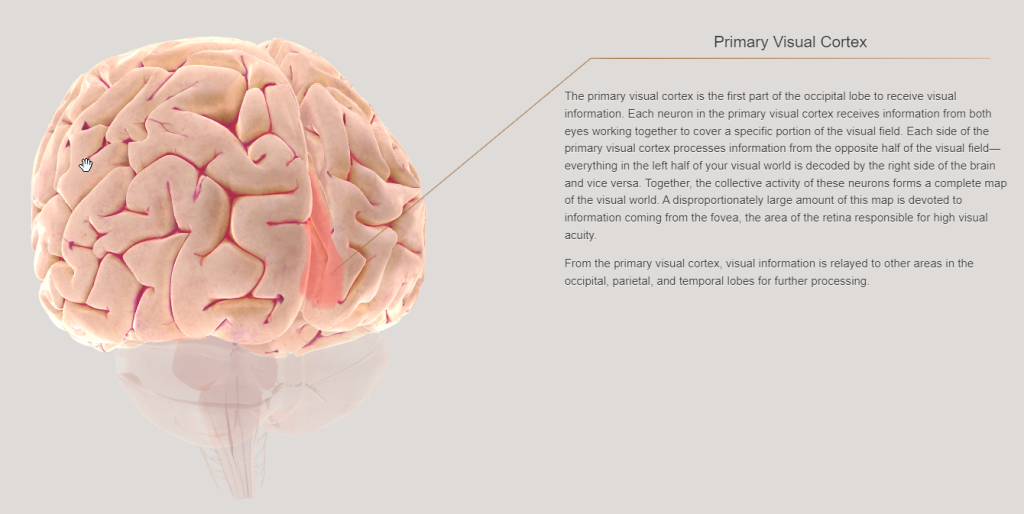
- occipital lobe, which is responsible for visual processing
- temporal Lobe, which is involved in auditory processing, as well as learning and memory.

Extension:
To learn more about the four lobes, including some visual information, you may want to visit the following resource: External brain anatomy.
One structure that forms part of the temporal lobe and that is frequently mentioned in the literature relevant to this eBook is the insula. The insula is involved in the anticipation of negative events, fear learning and avoidance behaviours (Davidson, 2010) It has also been found to play an important role in other types of emotional processing, e.g. in that of negative emotions such as disgust (Freberg, 2019). We will revisit the role of the insula when we look at decision-making in Chapter 2.
Extension:
For some 3D animation of the various lobes, visit BrainFacts and select each lobe in the dropdown menu on the left.
Thalamus and Hypothalamus
Both the thalamus and hypothalamus play an important role in emotional experiences (LeDoux, 1996; Papez, 1937, cited in Gluck et al., 2020), which is why we will look at them in more detail again in Chapter 3.
The thalamus can be referred to as the “gateway to the cortex” because it receives and processes sensory and regulatory input from the environment (with the exception of olfaction). The thalamus filters the information depending on a person’s arousal state before forwarding the filtered information to the cerebral cortex for further processing. The thalamus receives information from many brain regions, including the amygdala (discussed in more detail below) and sends projections back to these structures to create circuits involved in many different functions. The information passing through the thalamus relates, for example, to motivation, behaviour planning and sleep regulation. The thalamus also participates in learning and memory.

The hypothalamus plays an important role in regulating the autonomic nervous system. It connects to sites of the endocrine system that are involved in the release of hormones, including the adrenal glands, which are critical for the release of so-called “stress hormones”. The hypothalamus is also involved in regulating behaviours that are critical to keep us alive, including eating, drinking and temperature control.
Basal Ganglia
The basal ganglia (BG) consist of a group of brain structures including the subthalamic nucleus, the nucleus accumbens (sometimes called ventral striatum) and the corpus striatum, which again consists of the caudate nucleus, the putamen and the globus pallidus. You don’t have to remember all the substructures of the BG and their functions. You are introduced to these terms here because they may appear in some readings relating to the neuroscience of conflict management. You can then refer back to this chapter to make sense of any relevant readings. The BG are primarily involved in motor control and movement but have also been found to be involved in the perception and interpretation of emotions (Pierce & Péron, 2020) and emotional speech processing (Paulmann et al., 2011), which is why we are mentioning the basal ganglia here. The nucleus accumbens, one of the substructures of the BG that we mentioned above, is the region in which dopamine is released and research suggests a link between the nucleus accumbens and positive arousal, as well as behaviours associated with that arousal (Davidson, 2010).
Limbic System
The term “limbic system” was first introduced in the middle of the 20th century by MacLean (1952) and is still commonly used to describe several structures that are located on the inside edge of the cerebral cortex and that are thought to play a significant role in motivation, memory and emotion.
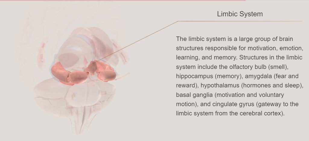
There has been ongoing disagreement as to which structures exactly form part of the limbic system (Ledoux & Phelps, 2008). Furthermore, neuroscience research has found that emotional processing cannot be localised to the structures of the limbic system (Feldman Barrett, 2017a; Feldman Barrett, 2020; Ledoux & Phelps, 2008; Pessoa, 2017). Research has also found that some structures of the limbic system, e.g., the hippocampus, are far more important for other cognitive processes (e.g., memory) than for processes involved in the experience of emotions (Ledoux & Phelps, 2008). Furthermore, multiple brain areas, of which many are not included in the limbic system, have been found to play an important role in the perception, experience and communication of emotions (Feldman Barrett, 2017a; Feldman Barrett, 2020; Ledoux & Phelps, 2008; Pessoa, 2017). Several contemporary neuroscientists reject both the idea that there is an ancient, emotional centre in our brain as which the limbic system is frequently presented in both scientific publications and popular press (and surely you would have come across courses, books or articles that promised you to help you learn to control your emotional brain), and the idea that emotions and cognitive processes can be clearly separated (Feldman Barrett, 2017a; Feldman Barrett, 2020; LeDoux & Brown, 2017; Ledoux & Phelps, 2008; Lempert & Phelps, 2016).
While we acknowledge that the experience of emotions is not limited to the limbic system, we will consider the limbic system here as the umbrella term that typically refers to several brain structures that play an important role in emotional experiences. Many sources include at a minimum the hippocampus, amygdala and cingulate cortex, which we will introduce below in more detail. You may wish to watch this short video as an introduction to these structures [1:52]:
Hippocampus
The hippocampus is located inside the temporal lobe and plays an important role in learning and memory, especially for episodic memory and autobiographical events (note how these functions relate much more to cognition than to emotions) (Gluck et al., 2020). The hippocampus is vulnerable to damage caused by the experience of stress. People who are exposed to ongoing conflict may suffer chronic stress and are thus vulnerable to hippocampus damage, which may again result in the loss of memory.
Amygdala
The amygdala has been found to play an important role in emotional memories, identifying emotional stimuli, including the emotional stage of other people, and physical arousal in response to certain stimuli (Davidson, 2010; Freberg, 2019). The amygdala is particularly active when we are confronted with danger/ thtrets and when we are in negative emotional states, but it also appears to play a role in reward and some positive emotional states (Freberg, 2019; Ledoux & Phelps, 2008). Furthermore, research has found that the amygdala also reacts to emotionally neutral, but unusual stimuli, which may suggest that the amygdala processes novel, unusual stimuli, especially those that might be important to safety and survival (Freberg, 2019; Ledoux & Phelps, 2008). We will look at the amygdala in much more detail in Chapter 3 since this structure has been found to play an important role in the perception and experience of emotions (as well as during “the stress response”). If you want to learn a bit more about the amygdala as early as now, please watch this video:
Cingulate Cortex
The cingulate cortex consists of the anterior cingulate cortex (ACC) and posterior cingulate cortex (PCC). You may come across both terms in readings on neuroscience and conflict management, which is why they are both briefly mentioned here. The ACC is involved in emotion regulation and decision-making, while the PCC is of interest for its involvement in memory and consciousness.