6 Muscular Case Studies
Chapter Objectives
- Describe the anatomical variation, population incidence of a range of phenotypes, and Australian and New Zealand context of select examples of the muscular system, including the:
- biceps brachii
- Discuss the clinical and/or professional impact of anatomical variation of the muscular system on patients and/or professional settings
Biceps Brachii
Variation Classification: Morphology and Frequency
Description of Anatomy
The biceps brachii is located in the anterior compartment of the brachium, and as its’ name suggests (‘bi’ = two; ‘ceps’ = heads), most commonly consists of two heads. The long and short heads of the biceps brachii attach from the supraglenoid tubercle and coracoid process of the scapula, respectively. The two heads then insert via a common tendon onto the radial tuberosity with an additional medial fascial slip, the bicipital aponeurosis inserting onto the anteromedial muscles of the antebrachium.
Examples of Phenotypic Variation
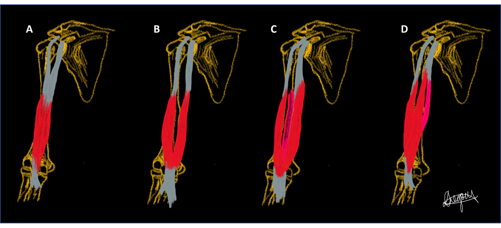
The biceps brachii can display supernumerary (accessory) heads with up to seven heads being reported and morphological variations in its attachment sites (both the origin and insertion). Supernumerary heads are most often observed unilaterally. In some cases the biceps brachii demonstrates a third head which most commonly arises from the shaft of the humerus distal to the insertion of the coracobrachialis (frequency variation). It has been hypothesised that supernumerary muscles in the upper limb may be an evolutionary trait, with muscles being more simple and slender in humans compared to primates and the cases of supernumerary heads decreasing over time demonstrating secular trends in incidence of this variation.
Four phenotypes of the biceps brachii are presented in a series of diagrams below. Use the slider tool to move between each phenotype and read the text underneath that describes each phenotype. By dragging the slider tool you can view the transition between each phenotype to clearly see how each phenotype differs from the next.
Population Frequencies of Phenotypic Variation of Supernumerary heads
| Source | Szewczyk et al. (2022) | Rodríguez-Niedenführ (2003) | Ballesteros et al. (2013) | Cucca et al. (2010) | Al-Kushi (2013) | Kosugi et al. (1992) | Nayak et al. (2006) | |
| Population | Central European | UK | Colombia | Australia (Western Australia) | Saudi Arabia | Japan | India | |
| Sample size | 101 (202 arms)
(Cadaver) |
175 (350 arms) (Cadaver) |
53 (106 arms)
(Cadaver) |
20 arms
(Cadaver) |
20 (40 arms)
(Cadaver) |
273 (546 arms)
(Cadaver) |
48
(Cadaver) |
|
| 2 heads |
64% of cadavers
2.5%* Single muscle belly 29.2%* Two muscle bellies
|
84.6% of cadavers | 80.2%* | 80%*
30%* Two bellies |
85% | 78.8% | 97.9% | |
| 3 heads |
26% of cadavers | 13.1% of cadavers
9%* Inferomedial head 1.5%* Superior humeral head 0.3%* Inferolateral head |
19.8%* Inferomedial head | 20%*
15%* Inferomedial head 5%* Superior humeral head |
15%* | 21.2% | 2.1% | |
| 4 heads |
6% of cadavers | 2.9% of cadavers | 0% | 0% | 0% | 0% | ||
| 5 heads |
4% of cadavers | 0% | 0% | 0% | 0% | 0% | ||
*% of total arms
Australian and New Zealand Context
In 2009, Fogg et al., completed a cadaveric dissection study within an Australian population aimed to describe the morphology of the distal biceps brachii tendon, to inform the common surgical approach used for tendon rupture (endobutton repair). Authors dissected 46 embalmed cadaveric arms (M =16, F = 18) and identified a variable number of distinct tendinous bands of both heads of the biceps brachii enclosed within the tendinous sheath, as opposed to just the typically described two tendinous bands associated with the short and long head of the biceps respectively. The tendinous bands occurred in varying numbers (mean 4.5 ± 2), each with a distinct attachment to the radius from immediately proximal to the radial tuberosity to distal to the radial tuberosity. The exact point of attachment and the distance between each attachment point was highly varied. Identification and description of these bands within the tendinous sheath of the biceps brachii is important, as commonly the endobutton surgical repair utilises stitches that pass through only the margin of the tendon; leaving several tendon bands free to move proximally. Authors suggest that the use of sutures that cross the width of the tendon may improve surgical outcomes (Fogg et al., 2009).
Case Study on Biceps Brachii: Case of supernumerary head of biceps brachii
A 34 year old male patient presents to the emergency department following a motorcycle accident. Dislocation of the right and left glenohumeral joints and rotator cuff muscle tears are suspected. During assessment of the patient’s MRI of the shoulder, a third head of the right biceps brachii is observed.
QUESTION 1: Would a supernumerary head of the biceps brachii result in any functional advantage to the patient?
QUESTION 2: Why is an accessory head of the biceps brachii sometimes mistaken for a tumour during medical image interpretation?
QUESTION 3: Compare and contrast the anatomy of the anterior muscle compartment of the brachium between humans and primates, and hypothesise why supernumerary heads of the biceps brachii suggest an evolutionary trait. Refer to Oxnard and Franklin (2008) for guidance.
References and Further Reading
Oxnard, C. ., & Franklin, D. (2008). Ghosts of the Past II: Muscles and Fasciae in the Primate Forelimb Domain. Folia Primatologica, 79(6), 441–457. https://doi.org/10.1159/000151357
Rodríguez-Niedenführ, M., Vázquez, T., Choi, D., Parkin, I., & Sañudo, J. R. (2003). Supernumerary humeral heads of the biceps brachii muscle revisited. Clinical Anatomy (New York, N.Y.), 16(3), 197–203. https://doi.org/10.1002/ca.10060
Szewczyk, B., Sanudo, J. R., Podgórski, M., Zielinska, N., Pires, M. B., Aragonés, P., & Olewnik, Ł. (2022). A Proposal for a New Classification of the Supernumerary Heads of the Biceps Brachii Muscle. BioMed Research International, 2022, 1510363–1510369. https://doi.org/10.1155/2022/1510363
