1 Introduction to Anatomical Variation
Chapter Objectives
- Define human anatomical variation and acknowledge its prevalence.
- Recognise and describe examples of anatomical variation.
Definition of Anatomical Variation
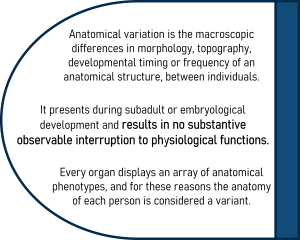
Anatomical variation is defined as the macroscopic differences in topography (location), morphology (shape and size), developmental timing or frequency (number) of an anatomical structure between individuals. It presents during subadult or embryological development and results in no substantive observable interruption to physiological function. Every organ displays an array of anatomical phenotypes and for this reason the anatomy of each person is considered a variant.
What this really means, is that the anatomy of each of us, is different! When learning anatomy, we are generally taught the ‘typical’ anatomy, or the most common anatomical phenotype, but it’s important for us, as health professionals, to understand that this anatomy isn’t the same for everyone. Therefore it’s vital that we are capable of recognising and describing different anatomical phenotypes.
Introduction to Anatomical Variation Video
Hotspot activity
The diagram below outlines just some examples of anatomical variation seen within the human body. Click on the ‘information’ icons to explore these examples to appreciate how common anatomical variation actually is!
[“Female shadow anatomy without labels” by Mikael Häggström/ Wikimedia Commons / Public domain]
Anatomical Variation Terminology
The language that we use to describe anatomical variation, and anatomy more generally, has adapted over the years to ensure inclusivity and recognition of the diversity of the human form. Andreas Vesalius, a sixteenth century anatomist, first described variant anatomy as ‘monstrous’ and ‘unnatural’ forms (Siraisi, 1994). Likely, this exclusionary view of anatomical variation, has influenced the way contemporary anatomists describe anatomical variants. For example, one more recent definition refers to variant anatomy as “deviations from the normal arrangement of an anatomical structure..” (Dangelo, 2004). The use of the term normal implies that anything that falls outside of normality is therefore considered abnormal – which is often associated with a negative or unwanted outcome. In an anatomy context, this is simply not the case! Just because an individual’s anatomy differs to what is typically described or isn’t described in a text book, does not mean that it is abnormal.
Instead of using the terms ‘normal’ and ‘abnormal’ when describing anatomy, we encourage you to use the word phenotype to describe the range of anatomy observed between individuals. Using terms such as most commonly described, frequent and typical anatomy, can also be useful in describing different phenotypic presentations. As an example; ‘the absence of an accessory bone within the foot is the most typically described anatomy, and the presence of an accessory bone is one other possible anatomical phenotype’ – as opposed to ‘the absence of an accessory bone within the foot is the normal anatomy, and the presence of an accessory bone is abnormal’.
The use of this inclusive terminology:
- Encourages acceptance of the diversity of all individuals.
- Aligns with the guidelines for inclusivity and cultural safety within health care.
- Broadens a health professional’s appreciation and recognition that anatomical variation is in fact normality.
Keep this terminology in mind as you move through the rest of this chapter – particularly when you practice describing different anatomical phenotypes.
References and further reading
Siraisi, N. G. (1994). Vesalius and Human Diversity in De humani corporis fabrica. Journal of the Warburg and Courtauld Institutes, 57(1), 60-88.
Dangelo JG. (2004). Anatomia humana sistêmica e segmentar. Anatomia Humana Sistêmicae Segmentar. p. 671-671.
Types of Anatomical Variation
There are four main categories of anatomical variation: topography or location of an organ or structure; morphology or shape, size and colour of an organ or structure; developmental timing of when the structure appears or matures; and frequency in appearance of a structure.
1. Topography
Variation in topography refers to differences in location of anatomical structures. The position or location of an organ can vary between one individual and another. Two examples of topographical variation are the locations of the vermiform appendix of the large intestine and the kidney’s.
Figure 1.1. This image displays the typical topography of the kidney’s (left) and an example of a pelvic kidney (right). [Case courtesy of Bruno Di Muzio, Radiopaedia.org, rID: 53683 & rID: 28076 CC BY-NC-SA 3.0]
Figure 1.2. This schematic shows the range of topography of the vermiform appendix. [Image: L. Gregory CC BY-NC 4.0. Adapted from “Grant 1962 172a” by John Charles Boileau Grant / Wikimedia Commons / Public Domain]
2. Morphology
Variation in morphology, refers to shape, size and colour differences in anatomy. There are many examples of morphological variations including our stomach shape, the shape and length of our bones and simply the size of our organs. Morphology also includes variations in colour. For example, our eye colour and skin colour!
Stomach morphology
Morphological variations in relation to the shape and size of the stomach are frequent between individuals. Most commonly, the stomach is classified as a J shape, whereby the body of the stomach is vertical (Figure 1.3). When the body of the stomach is orientated more obliquely or horizontally, it is classified as steer horn in shape. Another morphological variation of the stomach is the cascade classification. This classification describes the stomach when the fundus of the stomach is inverted – meaning that the fundus fills first, then overflows into the body of the stomach creating a cascading effect.

Pelvic morphology
The skeletal anatomy of the pelvis provides multiple examples of morphological variation, particularly when comparing the male and female pelvis (Figure 1.4). Some specific bony features of the pelvis which commonly display variation are the sub-pubic angle; the angle created by the left and right pubis bones, inferior to the pubic symphysis which is greater in females.

Sternum morphology
The sternum has a number of morphology variants. Explore the following two videos to delve into two phenotypes of the xiphoid process and an example of sternal foramen.
3. Developmental Timing
Developmental timing variants are variations in our anatomy that present as a consequence of varying rates of growth and development. Individuals of the same chronological age, can display different levels of development, resulting in significant anatomical variation within the population.
The development of the carpal bones, for example, involves ossification of cartilage to bone, occurring in a predictable sequence during subadult development. Differences in the carpal bone anatomy between individuals at different age groups, is an example of anatomical variation (Figure 1.5). Individual variability in the rate of growth is also common, and provides another example of anatomical variation whereby individuals of the same chronological age display different anatomical phenotypes as they are growing and developing at different rates (Figure 1.6).
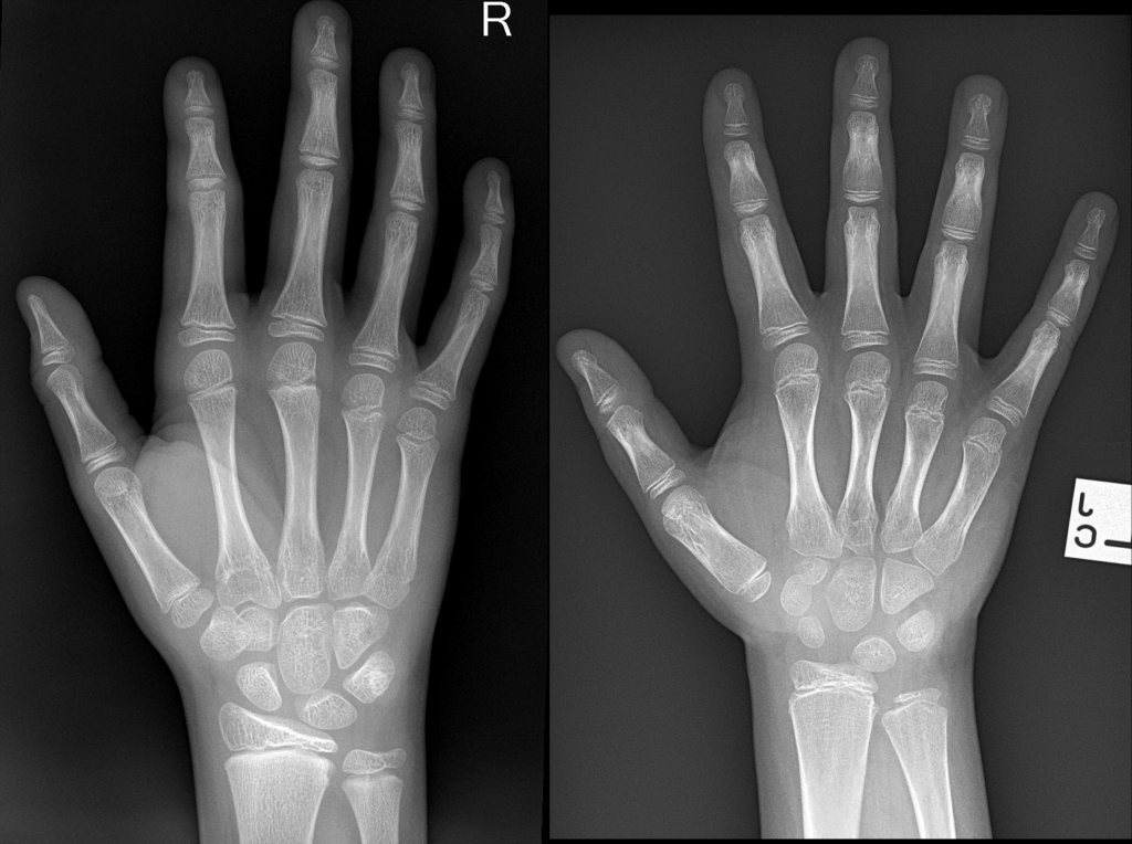
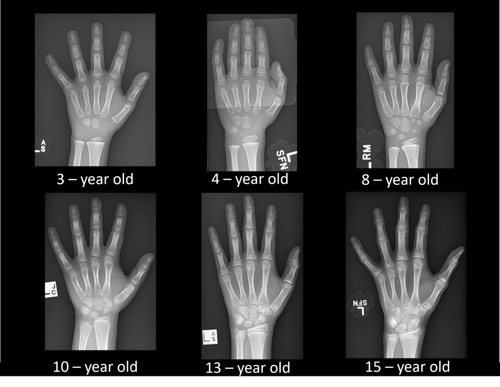
Figure 1.5 (left). This image compares the skeletal development of the hand of two different individuals of the same chronological age of ten. Variation in the rate of development of the carpal bones is visible. [Case courtesy of Calum Worsley and Dr Jeremy Jones, Radiopedia.org, rID: 93858 (left) & rID: 23244 (right) CC BY-NC-SA 3.0]
Figure 1.6 (right). Carpal bone development from three to fifteen years of age. [Case courtesy of Dr Jeremy Jones, Radiopedia.org, rID: 23244 CC BY-NC-SA 3.0]
4. Frequency
Frequency refers to variations in the number of times an anatomical structure appears. The presence of duplex (double) ureters, accessory bones in the hands and feet or the presentation of accessory muscles, are examples of variation in frequency.
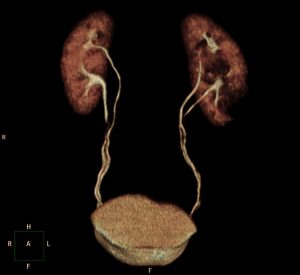
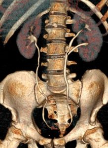
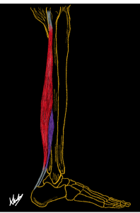
Figure 1.7. (left) A 3D rendered image displaying a duplex collecting system [Case courtesy of Dr David Cuete, Radiopedia.org, rID: 27556 CC BY-NC-SA 3.0]
Figure 1.8 (middle). A 3D rendered image displaying duplex left kidney with a bifid left ureter [Case courtesy of Dr Roberto Schubert, Radiopedia.org, rID: 15788 CC BY-NC-SA 3.0]
Figure 1.9 (right). Diagrammatic representation of an accessory soleus muscle (purple) and soleus (red). [Image: A. Kimmorley CC BY-NC 4.0]
Explore the following two videos to delve into some examples of frequency variants in the foot!
Combination of types
It is important to note that anatomical variations can also include multiple features across the different types of variations. For example, anatomical variants of the lung present in many forms including the presence of accessory fissures and lobes as well as the depth and completeness of the fissures itself. The computed tomography (CT) image below of the lung (Figure 1.10), displays a common variant in lung anatomy; the presence of a superior accessory fissure (indicated by the thin white line). This accessory fissure, is a variation in frequency, however it consequently alters the lung morphology as well by defining the boundary of an accessory lobe.
Figure 1.10. Computed tomography images of the typically described lung fissure anatomy (first image) and an example of an accessory fissure (second image).
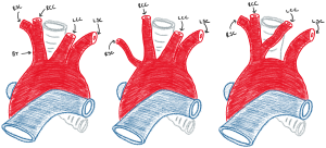
The aortic arch is another example where multiple types of anatomical variation are present within a single anatomical phenotype. The typically described phenotypic presentation of the branching of the aortic arch includes the brachiocephalic trunk, the left common carotid artery and left subclavian artery. However, variations to this branching pattern are quite common with differences in the number of vessels, and subsequently the location and course of the vessels, possible. For example, the brachiocephalic trunk can be completely absent. The right common carotid, right subclavian and left common carotid arteries can all have a common origin, resulting in only two aortic arch branches. This results in variations in both frequency, as well as topography as the course of the vessels is varied. The surrounding anatomy can also be varied as a consequence of these variations of the aortic arch. For example, he absence of an artery, would alter the course of the surrounding vessels, to ensure that oxygenated blood is still carried to all areas of the body.
Test your knowledge
Part of being understanding the concepts of anatomical variation is your ability to recognise and describe the different anatomical phenotypes you can see.
Using the flip cards below, practice identifying and describing different anatomical phenotypes.
Timing of Appearance of Anatomical Variation
Development of anatomical variations
The second part of the definition of anatomical variation, states that anatomical variation presents during embryological or subadult development. This means that during these stages of development; in-utero, infancy, childhood, adolescence and early adulthood, the interruption or alteration of typical developmental patterns may result in the formation of an anatomical variant.
These variations in the pattern of development can be influenced by multiple factors including nutrition, socioeconomic status and geographical location. Understanding and recognising the timing of development of anatomical variations is important to ensure accuracy when distinguishing between anatomical variation, anatomy of the ageing process and pathology.
Using the boundary of subadult development allows us to distinguish between the process of development and ageing. Ageing is often associated with a degeneration of a mature structure, while development is just that; the development and formation of a structure which primarily occurs during embryological and subadult maturation.
For example, intracranial calcifications (Figure 1.12), which are often small asymptomatic calcifications of small areas of the brain, can occur during both subadult and adult life. Importantly, they are changes to an already mature structure rather than variations that occur during the development of the structure. Therefore, they would be considered not as anatomical variation, but rather variations as a consequence as the process of ageing or tissue changes.
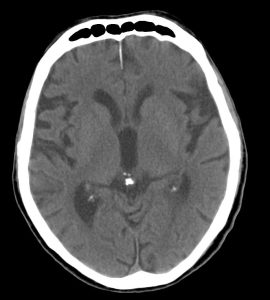
Figure 1.12 A computed tomography (CT) image of the head containing calcifications, which appear as small white areas on the image, in the location of the pineal gland and choroid plexus of the lateral ventricles. [Case courtesy of The Radswiki, Radiopedia.org, rID: 11770, CC-BY-NC-SA 3.0]
Anatomical Variation and Pathology
The third part of the definition of anatomical variation states that an anatomical variant results in no substantive, observable interruption to physiological function. This part of the definition enables us to distinguish between anatomical variation, pathology and congenital anomalies. Being able to distinguish between variation and pathology is important for health professionals when interpreting medical images and diagnosing patients.
When determining if an anatomical phenotype is a variant or pathology, three things need to be considered:
The following framework (Figure 1.13) can be used to help determine if the anatomical phenotype is an anatomical variant, pathology or a congenital anomaly.
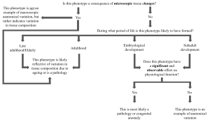
Figure 1.13. Anatomical variation decision tree.[Image: A. Kimmorley CC BY-NC 4.0]
Corpus Callosum morphology example
The corpus callosum is a C-shaped structure deep within the brain that functions to allow the two hemispheres of the brain to communicate to one another. Corpus callosum morphology can vary slightly between individuals, with larger morphological changes, such as partial agenesis (absence) of the corpus callosum also possible.
Consider whether the phenotype of partial agenesis of the corpus callosum is an example of anatomical variation, or pathology.
Figure 1.14: Normal Brain (MRI) [Left: Case courtesy of Assoc. Prof Frank Gaillard, Radiopaedia.org, rID: 37605 CC-BY-NC-SA 3.0.]
Figure 1.15: Partial agenesis of the corpus callosum in Down syndrome [Right: Case courtesy of Dr Ammar Haouimi, Radiopaedia.org, rID: 87829 CC-BY-NC-SA 3.0]
When would the anatomical phenotype develop?
Both slight and significant morphological changes of the corpus callosum can develop throughout embryological and/or subadult development – prior to the completion of callosal development.
Would function be significantly and visibly impaired?
Slight variations in corpus callosum morphology (shape and size) are extremely common and has no visible impact on physiological function. Therefore, these slight variations in morphology are considered anatomical variants.
A severe morphology variation such as partial or complete agenesis, most commonly interrupts the typical physiological function and visibly impairs an individual’s ability to function – growth may be impeded, they may have an inability to communicate or have vision and/or auditory loss. This type of interruption to function would be observable and substantive and therefore this morphology alteration would be classified as a congenital anomaly, rather than an anatomical variant.
Explore the following two videos to delve into two more examples of how we can distinguish between examples of anatomical variation and pathology.
Test your knowledge
Complete the following quiz to check your understanding of this module
