Imaging tests
John Perkins
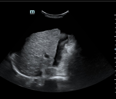
Abdominal ultrasound (liver, gallbladder) - handout available

Findings: Right hepatic lobe reduced in size with relative enlargement of the caudate lobe (C/RL ratio = 0.73). Liver surface nodular. Liver parenchyma shows increased echogenicity and coarse echotexture, indicative of diffuse fatty infiltration. Noted periportal oedema with perihilar lymphadenopathy. Dilated portal vein (15mm), low portal venous velocity, biphasic venous flow. No focal hepatic lesions. Abdominal ascites noted. Gallbladder and biliary ducts appear normal. Mild splenomegaly noted.
Impression: Severe chronic liver disease.
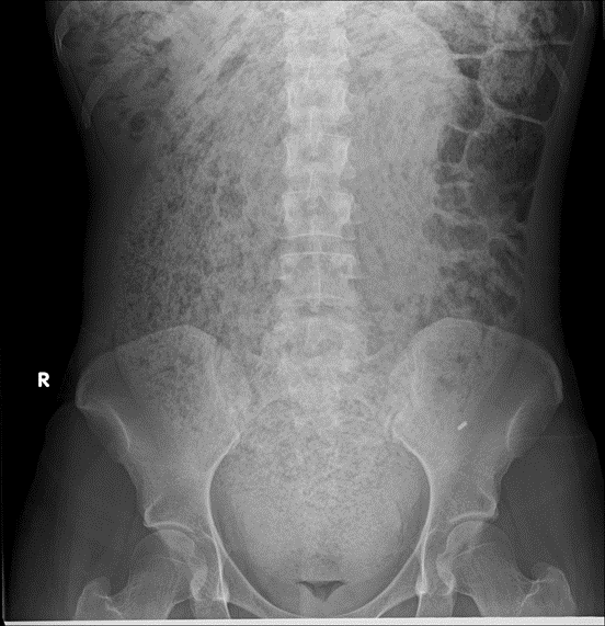
Abdominal x-ray - handout available

Interpretation. Do not provide to student/team unless requested in consultation.
Phenomenal amount of faecal loading throughout the abdomen with a markedly distended rectum. Minimal air in the distal rectum. Consistent with severe constipation.
Other imaging
| Investigation | Case result |
|---|---|
| Barium Enema | Not indicated |
| Barium Swallow | Not indicated |
| CT Abdomen/Pelvis | Abdominopelvic ascites. Liver surface nodular, with parenchymal heterogeneity and lobar atrophy. Signs of portal hypertension, periportal oedema and lymphadenopathy. Mild splenomegaly. |
| CT Chest (High Resolution) | Normal |
| CT Coronary Angiogram | Normal |
| CT Head | Normal |
| CT KUB (Kidneys, Ureters, Bladder) | Normal |
| CT Pulmonary Angiogram | Normal |
| Echocardiogram | Normal |
| Mammogram | Not indicated |
| MRI (General) | Normal |
| MRI Brain | Normal |
| Nuclear Medicine | Normal |
| Ultrasound Scan | See above (Liver Ultrasound) |
| X-Ray Abdomen | See above |
| X-Ray Chest | Normal |
| X-Ray Orthopaedic (Hands/Knees/Bone Age) | Normal |

