Case 7 – Abdominal US (liver, gallbladder) – Handout
John Perkins
Abdominal ultrasound

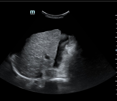
Findings: Right hepatic lobe reduced in size with relative enlargement of the caudate lobe (C/RL ratio = 0.73). Liver surface nodular. Liver parenchyma shows increased echogenicity and coarse echotexture, indicative of diffuse fatty infiltration. Noted periportal oedema with perihilar lymphadenopathy. Dilated portal vein (15mm), low portal venous velocity, biphasic venous flow. No focal hepatic lesions. Abdominal ascites noted. Gallbladder and biliary ducts appear normal. Mild splenomegaly noted.
Impression: Severe chronic liver disease.

