Imaging tests
Bruce Adamski
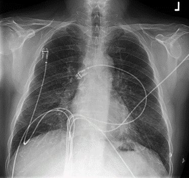
Chest x-ray - handout available

Interpretation. Do not provide to student/team unless requested in consultation.
Kerley B lines. ECG leads in place. Otherwise normal.
CT coronary angiogram - handout available
Scattered atherosclerotic plaque.
Left coronary artery is not opacified in its course and is thickened. Myocardial hypoperfusion can be seen in left anterior descending artery (LAD) perfusion bed. Right coronary artery appears partially stenosed in mid to distal course.
No pericardial effusion.
Other imaging
| Investigation | Case result |
|---|---|
| Barium Enema | Normal |
| Barium Swallow | Normal |
| CT Abdomen/Pelvis | Normal |
| CT Chest (High Resolution) | Ground glass infiltrate with smooth interlobular septal thickening and peribronchial cuffing consistent with early heart failure. |
| CT Coronary Angiogram | See above |
| CT Head | Normal |
| CT KUB (Kidneys, Ureters, Bladder) | Normal |
| CT Pulmonary Angiogram | Normal |
| Echocardiogram | Ejection Fraction 40%. Anterolateral wall hypokinesia. No effusion. Normal Valve function |
| Mammogram | Not indicated |
| MRI (General) | Normal |
| MRI Brain | Normal |
| Nuclear Medicine / Myocardial perfusion | Severe fixed perfusion defect in the anterior, lateral, and inferior wall of the left ventricle, and interventricular septum. Mildly reduced right ventricular perfusion. |
| Ultrasound Scan | Normal |
| X-Ray Abdomen | Normal |
| X-Ray Chest | See above |
| X-Ray Orthopaedic (Hands/Knees/Bone Age) | Normal |

