Imaging tests
Kyle Jennar
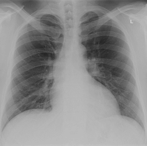
Chest x-ray - handout available

Interpretation. Do not provide to student/team unless requested in consultation. Lungs and pleural spaces are normal. Mild cardiomegaly..
Echocardiogram report - handout available
Left ventricular chamber size mildly dilated with increased wall thickness. Normal left ventricular systolic function with no evidence of regional wall motion abnormalities. Ejection fraction 65%.
Mitral valve appears normal in structure. Normal mobility of mitral leaflets. There is moderate 2+ mitral regurgitation.
Left atrium is enlarged in size, incompletely visualised. Small echogenic thrombus in left appendage.
Right ventricular chamber size normal with normal function. Right atrium normal in size. Tricuspid valve normal. Normal pericardium, no pericardial effusion.
Other imaging
| Investigation | Case result |
|---|---|
| Barium Enema | Normal |
| Barium Swallow | Normal |
| CT Abdomen/Pelvis | Hepatomegaly. Diffuse hepatosteatosis, increased nodularity. |
| CT Chest (High Resolution) | Cardiomegaly. Thrombus in left atrial appendage. |
| CT Coronary Angiogram | Normal |
| CT Head | Normal |
| CT KUB (Kidneys, Ureters, Bladder) | Normal |
| CT Pulmonary Angiogram | Normal |
| Echocardiogram | See above |
| Mammogram | Normal |
| MRI (General) | Normal |
| MRI Brain | Normal |
| Nuclear Medicine | Normal |
| Ultrasound Scan | Normal |
| X-Ray Abdomen | Normal |
| X-Ray Chest | See above |
| X-Ray Orthopaedic (Hands/Knees/Bone Age) | Normal |

