Imaging tests
Olive Greene
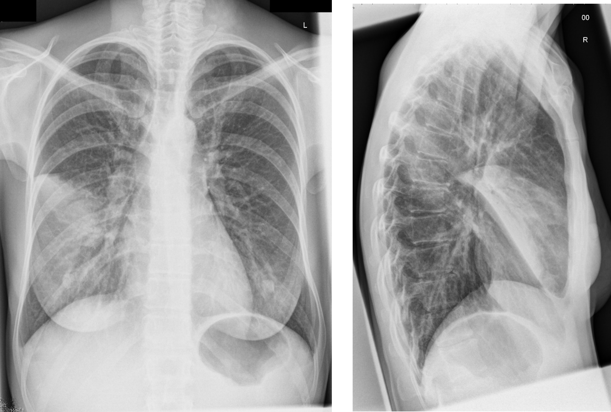
Chest x-ray - handout available

Interpretation. Do not provide to student/team unless requested in consultation.
Right middle lobe consolidation. Likely of infectious origin. Clinical correlation required.
Other imaging
| Investigation | Case result |
|---|---|
| Barium Enema | Normal |
| Barium Swallow | Normal |
| CT Abdomen/Pelvis | Normal |
| CT Chest (High Resolution) | Right middle lobe consolidation, with mild related pleural thickening, and minimal effusion in right pleural cavity. Widespread mild peribronchial inflammation. Heart size is normal. No pericardial effusion. The mediastinum structures have normal configuration. Chest wall is unremarkable. |
| CT Coronary Angiogram | Normal |
| CT Head | Normal |
| CT KUB (Kidneys, Ureters, Bladder) | Normal |
| CT Pulmonary Angiogram | Normal |
| Echocardiogram | Normal |
| Mammogram | Normal |
| MRI (General) | Normal |
| MRI Brain | Normal |
| Nuclear Medicine | Not indicated |
| Ultrasound Scan | Normal |
| X-Ray Abdomen | Normal |
| X-Ray Chest | See above |
| X-Ray Orthopaedic (Hands/Knees/Bone Age) | Normal |

