Imaging tests
Mike Simpson
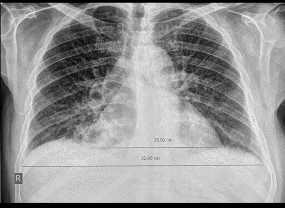
Chest x-ray - handout available

Interpretation. Do not provide to student/team unless requested in consultation.
Enlarged cardiac silhouette. Increased hilar markings with upper lobe diversion. No pleural effusions.
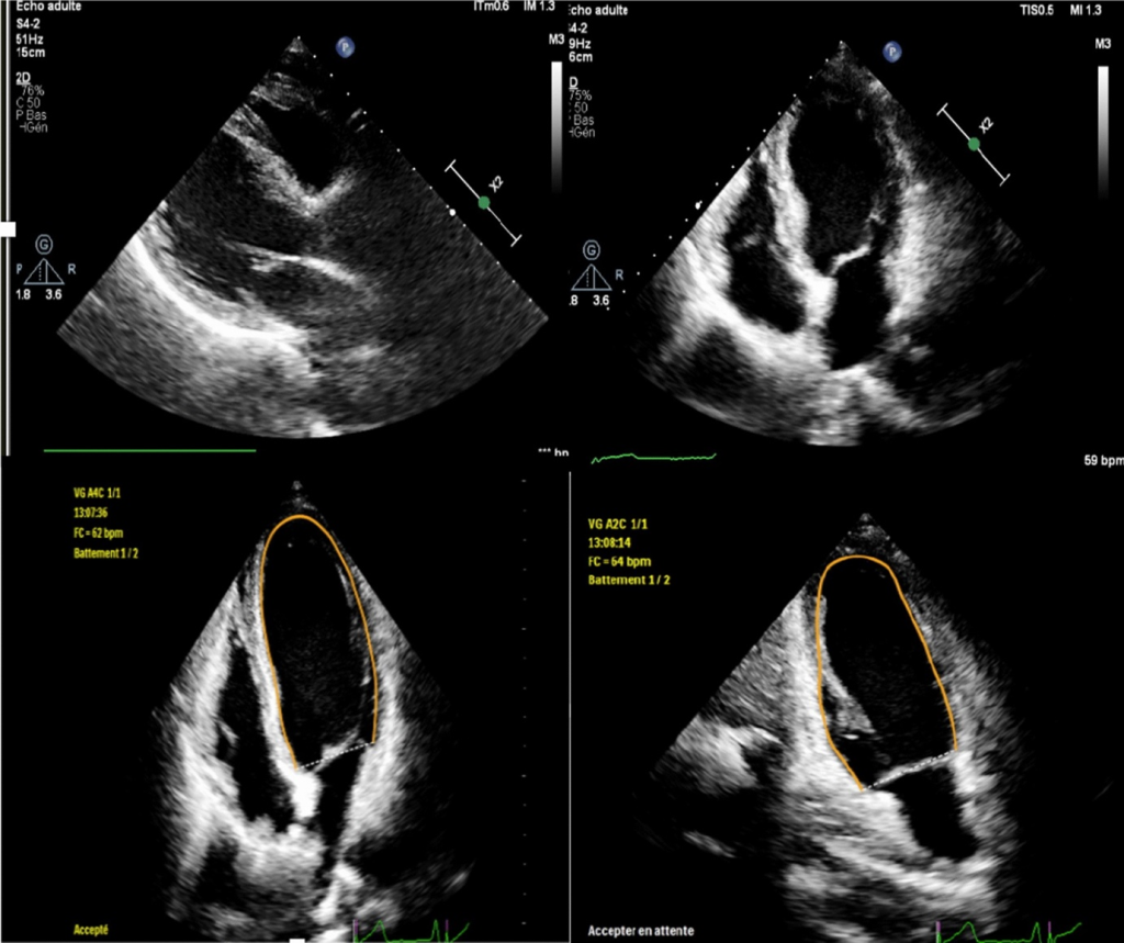
Echocardiogram report - handout available

Nil valvular disease. Mild-moderate right ventricular dysfunction, significant left ventricular dysfunction. Left Ventricle Ejection Fraction 30%. Left ventricular dilatation and mild hypokinesis noted.
Other imaging
| Investigation | Case result |
|---|---|
| Barium Enema | Normal |
| Barium Swallow | Normal |
| CT Abdomen/Pelvis | Normal |
| CT Chest (High Resolution) | Interstitial pulmonary oedema. Cardiomegaly. |
| CT Coronary Angiogram | Normal |
| CT Head | Normal |
| CT KUB (Kidneys, Ureters, Bladder) | Normal |
| CT Pulmonary Angiogram | Excludes pulmonary embolism |
| Echocardiogram | See above |
| Mammogram | Normal |
| MRI (General) | Normal |
| MRI Brain | Normal |
| Nuclear Medicine / myocardial perfusion scan | Not indicated |
| Ultrasound Scan | Bilateral carotid Doppler ultrasound – 60% occlusion left carotid artery, 45% occlusion right carotid artery at the level of the carotid bifurcation. |
| X-Ray Abdomen | Normal |
| X-Ray Chest | See above |
| X-Ray Orthopaedic (Hands/Knees/Bone Age) | Normal |

