Case 5 – CT pulmonary angiogram – Handout
Payne Stark
CT pulmonary angiogram

Report
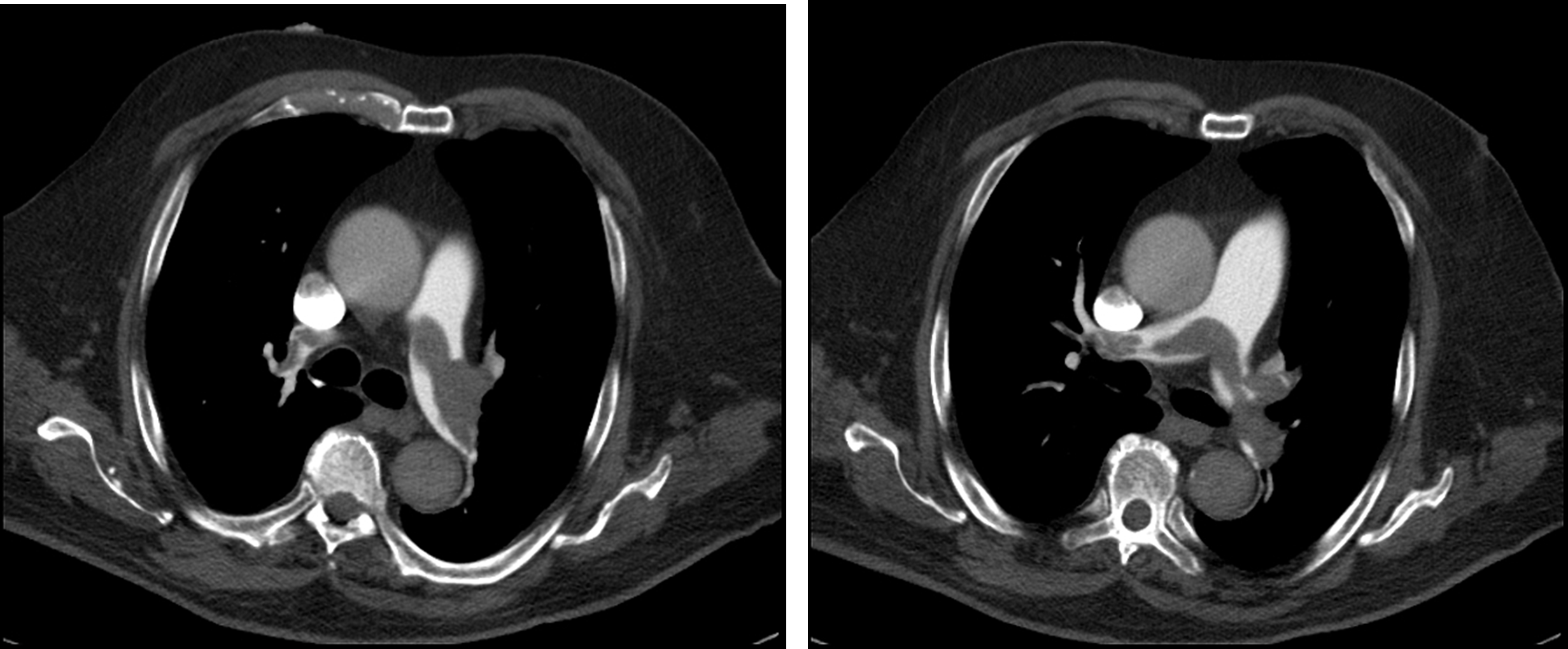
Large saddle embolus extending into the main pulmonary arteries, lobar and segmental arteries bilaterally. The lobar right lower lobe pulmonary artery is completely occluded, and within this distribution peripherally in the right lower lobe are multiple areas of peripheral parenchymal consolidation consistent with pulmonary infarcts (some of these are wedge-shaped). No effusion or pneumothorax. Tracheobronchial tree is patent. Mildly enlarged pulmonary trunk. Enlargement of the right ventricle, with bowing of the interventricular septum concerning representing right heart strain.
Impression
Large saddle pulmonary embolism with associated RLL pulmonary infarcts and evidence of right heart strain.

